Une augmentation du volume d’os par une greffe osseuse permet une plus grande stabilité et pérennité de l’implant
Les greffes osseuses préimplantaires sont indiquées lorsque la pose d’implants dentaires est impossible du fait d’un manque de volume osseux suffisant. Il existe plusieurs types de techniques de greffes osseuses pré-implantaires
NOS SOLUTIONS POUR UNE AUGMENTATION DU VOLUME OSSEUX
C'EST LE PRÉREQUIS DE LA PÉRENNITÉ DES IMPLANTS
LES GREFFES OSSEUSES PRÉ-IMPLANTAIRES
QU’EST CE QU’UNE GREFFE OSSEUSE PRÉIMPLANTAIRE ?
Les greffes osseuses pré-implantaires sont des interventions qui ont pour but d’augmenter le volume osseux lorsque ce dernier est insuffisant pour poser un ou des implants dentaires dans des conditions optimales Une augmentation du volume d’os alvéolaire par une greffe osseuse permet une plus grande stabilité et pérennité de l’implant.
Pourquoi le volume d’os alvéolaire est insuffisant ?
A la suite de la perte ou l’extraction d’une dent, une résorption osseuse physiologique et/ou pathologique survient systématiquement. Le volume d’os alvéolaire (c’est à dire la part d’os du maxillaire qui entoure, soutient et retient les dents) est dit insuffisant lorsqu’il ne permet plus de poser un ou des implant(s) dentaire(s) dans de bonnes conditions de longueur, de diamètre et de nombre.
C’est pour cela qu’il est conseillé de poser un implant dentaire immédiatement après l’extraction d’une dent lorsque cela est possible : l’os alvéolaire natif n’aura ainsi pas le temps de se résorber.
Explication : Lorsque la physiologie masticatrice des dents est normale, l’os alvéolaire maintient son volume grâce à des mécanismes spontanés de régénération osseuse.
Lors de la perte ou de l’extraction d’une dent, l’os alvéolaire qui entoure les racines des dents, diminue peu à peu en volume (largeur, épaisseur et hauteur) de 40 à 60%, durant les trois années qui suivent. Il est donc avantageux de réaliser une pose d’implant dentaire au plus vite, après la perte des dents.
Si une dent a été extraite il y a longtemps, alors le volume osseux est souvent déjà trop résorbé pour poser un implant dans des conditions optimales. Plus le nombre de dents augmente plus ce phénomène de résorption s’amplifie.
Si une dent est tombée spontanément c’est évidemment parce que la structure osseuse qui la retient à disparu partiellement ou totalement. Par définition, le volume osseux sera diminué et éventuellement insuffisant pour poser des implants dentaires.
Pour tous ces cas de figure, la solution est de procéder à une greffe osseuse préimplantaire afin de rétablir le volume osseux optimal pour poser un implant dentaire dans de bonnes conditions physiologiques.
LA GREFFE OSSEUSE :
UNE ÉTAPE INCONTOURNABLE POUR POSER DES IMPLANTS DENTAIRES?

Dans quels cas les greffes osseuses préimplantaires sont-elles indiquées?
Les greffes osseuses préimplantaires sont réalisées sur les patients ayant un volume osseux insuffisant d’os alvéolaire pour effectuer une pose d’implant dentaire dans de bonnes conditions mécaniques et physiologiques.
La décision de greffer de l’os est prise suite au diagnostic d’insuffisance de volume osseux implantaire. Ce diagnostic est posé selon les résultats de l’exploration du volume d’os alvéolaire disponible réalisée grâce à un scanner 3D. Cet examen effectué grâce au scanner cone beam 3D est indispensable pour mesurer précisément la quantité d’os dans la zone qui accueillera l’implant dentaire. De plus, il est aisé de vérifier en même temps s’il y a la présence d’un obstacle chirurgical.
Les procédures chirurgicales de greffes d’augmentation du volume osseux sont obligatoires dans les cas complexes d’implantologie, comme les poses d’implants après des extractions multiples et mise en charge immédiate.
POURQUOI RÉALISER UNE GREFFE OSSEUSE AVANT LA POSE D’UN IMPLANT DENTAIRE?
Il n’est pas toujours possible de poser un implant dentaire directement. Les règles de l’implantologie impliquent que les implants dentaires intra-osseux soient posés dans un volume d’os alvéolaire suffisant pour permettre une bonne cicatrisation et une vascularisation de l’os afin de diminuer le risque de péri-implantite. Cela implique que la crête alvéolaire des maxillaires dans laquelle sont posés les implants doit avoir un volume suffisant d’os disponible implantable. De même, la qualité de l’os est primordiale : un os peu dense, gras ou peu vascularisé seront des contre-indications à la pose d’implants dentaires. L’os alvéolaire qui reçoit l’implant dentaire doit fournir un ancrage efficace aux implants afin de garantir la pérennité de la prothèse dentaire implanto-portée.
Or, l’os alvéolaire peux avoir une hauteur ou une largeur insuffisante :
- Soit parce que le capital osseux chez certain patient est naturellement insuffisant avec un morphotype fin.
- Soit parce que l’os s’est résorbé, physiologiquement ou pathologiquement, suite aux extractions des dents (voir le chapitre sur les greffons osseux).
De plus, des structures anatomiques peuvent être des obstacles à la pose d’implants dentaires comme :
- La paire de nerf alvéolaire inférieure (NAI) qui parcourt la partie postérieure et horizontale de la mandibule à droite et à gauche.
- La paire de sinus maxillaire au-dessus de la partie basale postérieure du maxillaire et dans l’os de la pommette ou (os zygomatique) à droite et à gauche.
- Les fosses nasales au dessus de la partie antérieure du maxillaire au niveau bloc incisivo canin.
Donc, si la quantité et la qualité de l’os des crêtes osseuses implantables est devenue insuffisante, il est nécessaire d’augmenter son volume et sa densité grâce aux greffes osseuses et l’adjonction de facteurs de croissance contenu dans le Plasma Rich Platelet (PRP) et le Plasma Rich Fibrin (PRF).
C’est le domaine de la chirurgie pré-implantaire. Les différentes techniques chirurgicales à disposition pour augmenter le volume osseux disponible vont être discutées dans ce chapitre.
Les greffons osseux utilisés en chirurgie préimplantaire sont soit :
- Une greffe osseuse avec un prélèvement osseux sur le patient édenté. C’est une autogreffe osseuse puisqu’il est à la fois donneur et receveur.
- Une greffe osseuse sans prélèvement osseux avec un biomatériau comme greffon osseux. Le patient édenté est receveur mais pas donneur. Il peut s’agir d’un greffon allogène ou xénogène ou synthétique.

VOUS ÊTES UNIQUE !
CAS CLINIQUES DE RÉSOPTION OSSEUSE
RÉSULTATS CLINIQUES DES GREFFES OSSEUSES PRÉIMPLANTAIRES
QUELLES TYPES DE GREFFES OSSEUSES PRÉIMPLANTAIRES
ET POUR QUELLE RESTAURATION DE VOLUME?
Il existe plusieurs types de protocoles de greffes osseuses préimplantaires. Le choix de la technique dépend de la direction prépondérante du manque volumique. On distingue
- L’augmentation verticale lorsqu’il n’y a pas assez de hauteur d’os alvéolaire implantaire,
- L’augmentation horizontale lorsqu’il n’y pas assez d’épaisseur d’os alvéolaire implantaire
- Le comblement de l’alvéole dentaire après une ou plusieurs extractions dentaires simultanées pour restructurer le volume d’os alvéolaire implantaire dans toutes les directions.
En fonction du type de greffe effectuée, la technique chirurgicale de greffes osseuses préimplantaires peut avoir lieu soit au cabinet dentaire, ou soit en clinique, respectivement sous anesthésie locale ou générale.
Les petites interventions chirurgicales non invasives sont pour la plupart réalisées au cabinet dentaire sous sédation semi-inconsciente par voie intraveineuse avec l’assistance d’un médecin anesthésiste-réanimateur.
A la mâchoire supérieure:
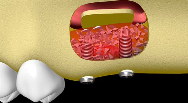
Suite à une résorption verticale de l’os alvéolaire postérieur de la mâchoire supérieure, la greffe osseuse de compensation du manque de hauteur d’os alvéolaire implantaire de référence est la technique de comblement des sinus ou sinus lift (élévation du plancher du sinus).
Ce protocole est indiqué dans les cas d’insuffisance osseuse en hauteur dans les zones postérieures du maxillaire supérieur sous le sinus.
La cavité aérienne que forme le sinus augmente de volume, par un phénomène de pneumatisation, lorsqu’une ou plusieurs molaires sont extraite et quelque fois les prémolaires. Le sinus gagne de la place au détriment de l’os alvéolaire disponible pour poser des implants, suite à la perte de ces dents. La partie basse du volume de la cavité aérienne du sinus doit être comblée avec un greffon osseux pour restaurer le volume d’os alvéolaire implantaire.
Ce protocole est choisi lorsque la hauteur résiduelle située sous le sinus ne dépasse pas 8 millimètres.
Dans certain cas de grosse résorption verticale la technique du sinus lift peut être associée à la technique de greffe d’apposition avec une matrice transvissée ou ROG (régénération osseuse guidée).
A la mâchoire inférieure :
Suite à une résorption verticale de l’os alvéolaire postérieur de la mâchoire inférieure, la greffe osseuse de compensation du manque de hauteur d’os alvéolaire implantaire de référence est la technique de greffe d’apposition avec une matrice transvissée ou ROG ( régénération osseuse guidée). Une grille métallique en titane est fixée par des vis à l’os et soulage le greffon de la pression des tissus mous sus-jacent afin de limiter la résorption du greffon, induite par cette pression pendant la cicatrisation.
Ce protocole est indiqué dans les cas d’insuffisance osseuse en hauteur dans les zones postérieures du maxillaire inférieur.
Ce protocole est choisi lorsque la hauteur résiduelle située au-dessus du nerf alvéolaire inférieur ne dépasse pas 8 millimètres.
Suite à une résorption horizontale de l’os alvéolaire postérieur ou antérieur des mâchoires inférieures ou supérieures, la greffe osseuse de compensation du manque d’épaisseur d’os alvéolaire implantaire de référence est la technique de greffe d’apposition avec une matrice transvissée ou ROG (régénération osseuse guidée). Une grille métallique en titane est fixée par des vis d’ostéosynthèse sur le pourtour de la lacune d’os alvéolaire. Cette grille guide l’ostéosynthèse et soulage le greffon osseux de la pression des tissus mous. On sait que cette pression contrecarre la néo angiogénèse. Donc, limiter la pression sur le greffon équivaut à favoriser la fabrication d’un nouveau réseau vasculaire et par conséquent limiter la résorption du greffon induite par cette pression pendant la cicatrisation.
Ce type de greffe est réalisée sur les parois verticales externes des maxillaires supérieurs et inférieurs lorsque l’os n’est pas assez épais.
Pour autant les techniques de greffes horizontales sont variées:
- Régénération Osseuses Guidée (ROG),
- Ostéotomies segmentaires,
- Autogreffe osseuse d’apposition avec fixation et ostéosynthèse
- Ostéotomies longitudinales d’expansion.

LES DEUX GRANDES TECHNIQUES DE GREFFE OSSEUSE

Les greffes osseuses préimplantaires avec un prélèvement osseux sur le patient
Il s’agit d’une greffe dite autologue ou autogreffe : le chirurgien vient prélever sur le patient le greffon
- En extra buccale : sur l’os pariétal crânien, sur l’os iliaque, sur l’os huméral,
- En intra buccale : sur l’os du menton ou l’os ramique à la base inférieure de la mâchoire inférieure.
Dans ce type d’autogreffe, la technique chirurgicale va obligatoirement inclure deux temps opératoires et deux sites opératoires pour le prélèvement osseux et la fixation du greffon.
Les greffes osseuses préimplantaires sans prélèvement osseux sur le patient
Il existe d’autres types de greffons fabriqués et distribués par des laboratoires pharmaceutiques :
- Allogreffes: greffons issus de donneurs humains vivants volontaires ;
- xénogreffes: greffons d’origine animale ;
- alloplastiques ou biomatériaux synthétiques
Dans ce type de greffe qui fait intervenir des produits pharmaceutiques, la technique chirurgicale va inclure un temps opératoire et un site opératoire pour la fixation du greffon sans prélèvement osseux.
Le seul site externe à celui de la greffe est éventuellement celui de la prise de sang pour faire les PRF.

COMMENT SE PASSE L’OPÉRATION DE GREFFE OSSEUSE PRÉIMPLANTAIRE?

La greffe est réalisée sous anesthésie locale, avec l’aide ou non d’une sédation par intraveineuse. Il est également possible de la réaliser sous anesthésie générale.
Le chirurgien-dentiste pose le greffon, puis il l’immobilise avec différentes techniques en fonction du type de greffon. Cette greffe osseuse est parfois complétée par un apport de PRF (Plasma Riche en Fibrine).
Les implants dentaires pourront être posés :
- Soit immédiatement au moment de la greffe osseuse
- Soit quatre à six mois après l’intervention. Dans ce cas, avant l’implantation, le chirurgien-dentiste procède à un scanner pour vérifier si le volume osseux est optimal.
Les suites opératoires habituelles d’une greffe osseuse préimplantaire sont dépendantes de différents facteurs intrinsèques et extrinsèques : complexité, étendues, temps opératoire, lieu de l’intervention, présence d’une voie veineuse, type de greffon,
La greffe est une opération et il n’y a pas de petite chirurgie. La période de cicatrisation est une phase délicate ou différents aléas peuvent se produire : relâchement ou rupture des fils de suture, infection, inflammation non controlée, etc.
Durant cette période, il est possible qu’une douleur ou un gonflement surviennent.
Des complications apparaissent avec un taux très faible de moins de 10 % mais ils sont statistiquement inévitables.
Il est important de suivre certaines préconisations et contre-indications durant la cicatrisation :
- Le tabac est absolument proscrit un mois avant l’opération, et définitivement proscrit par la suite.
- Il est conseillé de dormir sur le dos, sans pression sur la joue du côté où la greffe a été réalisée pour éviter le déchirement des sutures. A cet effet le port d’un collier gonflable (type avion) est conseillé pour ne pas dormir sur le côté opéré et tirer sur les fils de suture.
- Les sports extrêmes sont proscrits, de même que les activités qui demandent des positions où la tête se retrouve vers le bas (yoga, arts martiaux, etc.)
- En cas de greffe osseuse sinusienne, il faut se moucher délicatement pendant les trois premières semaines.
- Une excellente hygiène bucco-dentaire est primordiale. Les bains de bouche sont interdits. Les gencives doivent être nettoyées à l’aide d’une brosse chirurgicale après chaque prise alimentaire.
- Durant les jours qui suivent l’opération, la nourriture ingérée doit être molle, froide ou tiède. Les aliments chauds, salés et acides sont à éviter.
Toute opération chirurgicale possède un risque de complications. En cas de doute, n’hésitez pas à contacter l’équipe chirurgicale.
Le risque le plus important est une infection du greffon qui peut provenir du sinus, ou d’une cicatrisation gingivale tardive. L’infection doit être éliminée par un traitement antibiotique, voire par un curetage de l’os. Le foyer infectieux peut mener à la perte d’un morceau du greffon, celle-ci pouvant nécessiter une nouvelle intervention.
Heureusement, le risque d’infection n’est pas répandu. Il ne concerne que 5 à 10% des opérations, et est observé en majorité chez les patients fumeurs, diabétiques, ou qui ne respectent pas une hygiène bucco-dentaire rigoureuse après l’intervention.


